Intern Report 8.28

internreport

Discussion by Mirjana Dimovska, MD

Case 1

45 year old female presents to the emergency department with intense bilateral hand pain after a glass etching arts and crafts project this morning. The patient states that she thinks she got some of the solution on the palms of her hands. On visual inspection, skin has corrugated appearance.

images

1. Which of the following electrolyte abnormality is most likely to lead to adverse effects in this patient?

A. hyponatermia

B. hypocalcemia

C. hypernatremia

D. hypokalemia

Case 2

A 56 year old dissolved male is brought into the emergency department via EMS. The patient smells strongly of alcohol and was noted to be sleeping on a metallic park bench during a thunderstorm. Upon exam, the patient has a feathering patterned burn to the right upper extremity and chest. GCS is 12 however patient is confused and unable to provide a history.

2. Which of the following physical exam findings is most likely?

A. Muscle necrosis distant to the site of injury

B. Compartment syndrome in RUE

C. Kissing burns

D. Tympanic membrane rupture

Case 3

A 3 year old child is brought in by his mother for mental status changes. The patient’s past medical history is significant for a recent emergency room visit for treatment of an extensive scald burn that the patient sustained while trying to lift a bowl of ramen noodles. On exam, the child is cyanotic, short of breath and lethargic. He has superficial scald burns on the left shoulder and upper back covered in ointment.

3. What is the most appropriate treatment plan?

A. Administration of methylene blue and supplemental O2

B. Contact child protective services immediately

C. Admit to burn service for debridement and pain control

D. Massive fluid resuscitation and supplemental O2

Bonus Question:

Which of the following does not fulfill criteria for transfer to a burn center?

A. 37 year old male pulled from a house fire with singed nose hairs and DIB

B. 15 year old female with partial thickness burns to the bilateral left upper and lower extremities

C. 6 year old male with full thickness burn to the right arm totaling <5% BSA

D. 52 year old female who sustained a superficial partial thickness grease burn to the right breast while cooking this morning.

Answers:

1. B. Hypocalcemia.

Hydrofluoric acid is a weak acid used in the petroleum industry, however also used for glass etching, as a rust remover and for the cleaning of cement and bricks. The free fluoride ion serves as a cation scavenger, notably for calcium and magnesium. The free fluoride ion can also inhibit the sodium potassium ATPase resulting in hyperkalemia. These electrolyte disturbances can result in QT prolongation, ventricular arrhythmias, hypotension, other badness. Hypocalcemia after a significant exposure is treated with IV calcium gluconate 10% or calcium chloride via central access and warrants admission to telemetry to monitor for arrhythmias. Burns as small as 2.5% BSA can be fatal in a high concentrated hydrofluoric acid exposure.

2. D. Tympanic membrane rupture.

This patient is victim to a lightning injury as evidenced by the feathering patterned burn. Blunt trauma associated with these injuries can result in skull fractures or cervical spine injuries. Consequently, tympanic membrane rupture is a common finding in these victims and can be secondary to a basilar skull fracture or may be the result of a shock wave or direct burn. Otoscopic exam is essential in suspected lightening injury patients as ossicular disruption can cause permanent hearing damage. Choices a-c are most consistent with an electrical injury.

3. A. Administration of methylene blue and supplemental O2.

This child is suffering from acquired methemoglobinemia precipitated by silver sulfadiazine cream. Decontamination with soap and water is recommended along with treatement with methylene blue 1-2mg/kg IV over 5 minutes for patients with symptomatic hypoxia or MethHgb levels >30%. Symptoms include difficulty in breathing, dysrhythmias, seizures, coma. Other adverse affects of silver sulfadiazine include hypersensitivity reactions, neutropenia, leucopenia.

Bonus: D. American Burn Association recommends transfer of the following to a burn center:

1. Partial thickness burns greater than 10% total body surface area (TBSA).

2. Burns that involve the face, hands, feet, genitalia, perineum, or major joints.

3. Third degree burns in any age group.

4. Electrical burns, including lightning injury.

5. Chemical burns.

6. Inhalation injury.

7. Burn injury in patients with preexisting medical disorders that could complicate management, prolong recovery, or affect mortality.

8. Any patient with burns and concomitant trauma (such as fractures) in which the burn injury poses the greatest risk of morbidity or mortality. In such cases, if the trauma poses the greater immediate risk, the patient may be initially stabilized in a trauma center before being transferred to a burn unit. Physician judgment will be necessary in such situations and should be in concert with the regional medical control plan and triage protocols.

9. Burned children in hospitals without qualified personnel or equipment for the care of children.

10. Burn injury in patients who will require special social, emotional, or rehabilitative intervention.

Intern Report 8.27

internreport

Case Discussion by Matt Ciejka, MD

HPI:

23 year-old female presents with complaint of intermittent left lower abdominal pain for the past 3 days. She also complains of occasional vaginal spotting that began 1 day prior. Her last menstrual period was 26 days ago. She denies any vaginal discharge otherwise. Her abdominal discomfort is characterized as a sharp sensation over the left lower abdomen, lasting for several minutes at a time. She endorses some associated nausea but no vomiting, diarrhea, nor blood in stools. She has not taken anything at home for her symptoms. She denies any lightheadedness or syncope. She has no other complaints at this time.

PMH: HTN, migraines, Graves’ disease

PSH: foot surgery

Meds: propranolol, PTU

Allergies: amoxicillin, Keflex, doxycycline, clindamycin, (all cause hives)

Social history: denies tobacco, alcohol, and illicit drugs

Family history: CHF, diabetes, HTN

EXAM:

Vitals: BP 142/79, HR 106, RR 18, T 36.6, SpO2 99% on RA

Gen: A/Ox3, NAD

HEENT: PERRL, TMs WNL, no rhinorrhea, no oropharyngeal erythema

CV: regular rhythm, tachycardic, no m/r/g

Resp: lungs CTAB, no respiratory distress

Abd: obese, soft, mild tenderness over left lower abdomen, no distension, no peritoneal signs

Musc: 5/5 strength in all extremities throughout

Skin: no rashes appreciated

Neuro: follows all commands, answers all questions appropriately, sensation intact throughout extremities

Pelvic: no vaginal discharge, scant blood in vaginal vault but no active bleeding, no cervical motion tenderness, no palpable adnexal masses, mild tenderness over left adnexal area, slightly enlarged soft uterus


Questions:

1) Which of the following is the most appropriate next test?

A. Abdominal x-ray

B. Abdominal/pelvic CT scan

C. Abdominal ultrasound

D. Urine human chorionic gonadotropin

E. Progesterone concentration


2) Which of the following is the most common etiology of ectopic pregnancy?

A. Previous medically-induced abortion

B. Previous tubal surgery

C. Intrauterine device (IUD) contraception use

D. History of pelvic inflammatory disease

E. In utero exposure to diethylstilbestrol (DES)


3) Which of the following combinations of ultrasound findings and blood work is most suggestive of an ectopic pregnancy?

A. Fluid in pouch of Douglas on ultrasound; serum progesterone 30 ng/mL

B. Absence of intrauterine gestational sac on transvaginal ultrasound; serum hCG 1,600 miU/mL

C. Absence of intrauterine gestational sac on transvaginal ultrasound; serum hCG 800 miU/mL

D. Absence of intrauterine gestational sac on transabdominal ultrasound; serum hCG 4,000 miU/mL

E. Absence of intrauterine gestational sac on transabdominal ultrasound; serum hCG 2,000 miU/mL

Answers:

1. (D) Female patients who are of reproductive age and present with complaints of abdominal pain and vaginal bleeding should initially receive a urine or serum pregnancy test. A qualitative urine hCG test is sensitive for detecting early pregnancy with thresholds as low as 10 mIU/mL to 100 mIU/mL, depending on the test brand. The test is 99% sensitive and 99% specific for pregnancy. If the urine hCG test is positive, one can initially perform transabdominal ultrasound examination to determine the location of the pregnancy and help rule out an ectopic. If an intrauterine pregnancy is not visualized, a transvaginal ultrasound examination can be performed. It should be noted that a serum progesterone level may help to ascertain whether or not a pregnancy is viable (>25 ng/mL suggests viability). If the urine hCG test is negative, one should consider other diagnoses such as PID, urinary tract infection or stone, gynecological issues such as fibroids or ovarian cysts, or GI issues such as diverticulitis or appendicitis. For evaluation of these issues, the other listed tests may be beneficial.
2. (D) The risk for ectopic increases secondary to mechanisms that affect the movement of a fertilized egg through the fallopian tube. Such mechanisms can be anatomical, such as tissue scarring, or functional, such as a decrease in fallopian tube motility. Pelvic inflammatory disease is the leading cause of ectopic pregnancy, and at least 50% of first ectopic pregnancies are associated with a history of PID. It is most often caused byN. gonorrheaor C. trachomatis, whose long-term untreated course can damage the structural integrity within fallopian tubes. Other risk factors for ectopic pregnancy include a prior ectopic pregnancy, endometriosis, and tubal and pelvic surgery by way of formed adhesions obstructing the fallopian tubes. Normal fallopian tube motility can also be impeded by hormonal imbalances involving progesterone. A pharmacological elevation of progesterone, such as from progesterone-only OCPs or IUDs is associated with ectopic pregnancy. In utero exposure to diethylstilbestrol (DES) has been shown to increase risk of ectopic pregnancy as well. A history of medically-induced abortion has not been shown to increase risk.
3. (B) The “discriminatory zone” is the range of serum hCG concentrations above which a gestational sac can be visualized consistently. Transabdominal ultrasound examination can consistently detect a gestational sac when the hCG level is greater than 6,500 mIU/mL. Absence of an intrauterine gestational sac on transabdominal ultrasound with hCG level greater than 6,500 is highly suggestive of an ectopic pregnancy. Transvaginal ultrasound is more sensitive for detection of intrauterine pregnancy and has a lower “discriminatory zone” than transabdominal ultrasound, as it can consistently detect intrauterine pregnancy in conjunction with a hCG level greater than 1,500 mIU/mL. Transvaginal ultrasonography with serum hCG level greater than 1,500 mIU/mL is 67-100% sensitive and 100% specific for detecting ectopic pregnancy. However, it must be noted that there is no hCG level at which the possibility of visible ectopic pregnancy can be ruled out with absolute certainty. Serum progesterone levels can identify patients at risk for ectopic pregnancy, although they are not diagnostic of ectopic pregnancy. Serum progesterone concentrations are higher in viable IUPs than in ectopic pregnancies or IUPs that are destined to abort. A progesterone level of 5 ng/mL or less indicates a nonviable pregnancy, such as ectopic or miscarriage, and excludes normal pregnancy with 100% sensitivity. Due to the poor reliability of progesterone levels in detecting ectopic pregnancy, however, serum hCG levels are used more often in conjunction with ultrasound.
SOURCES

http://www.aafp.org/afp/2005/1101/p1707.html

http://www.aafp.org/afp/2000/0215/p1080.html

http://www.uptodate.com/contents/ectopic-pregnancy-clinical-manifestations-and-diagnosis

Herbst AL, et al. Ectopic pregnancy. Comprehensive gynecology. 2nd ed. St. Louis: Mosby-Year Book; 1992:457–88

Malhotra N, et al. Operative Obstetrics and Gynecology. JP Medical Ltd 2014: 439-440


Intern Report 8.26


internreport

Case Presented by Barry Kang, MD

Questions:


1)
Patient is brought into resuscitation as a trauma code 1. He has been shot multiple times in the chest. He is intubated and is swept off to the operating room. As your are leaving the resuscitation bay a DPD officer approaches you and asks you about the patient and what is going on. What do you respond?

a) I’m sorry sir the only thing I can tell you is that the patient is in critical condition and that he is one his way to the operating room.
b) His name is John Brooks and he got shot multiple times in the chest and abdomen. He had to be intubated and is in critical condition. He was just taken to the operating room.
c) He is middle age African American male who has sustained multiple gunshot wounds and was just taken to the operating room.
d) Here is FIN number with his name, age and birthday. He has sustained multiple gunshot wounds to the chest and abdomen. He was just taken to the operating room.


2) You have just seen an interesting case in MOD 2 and the patient was just sent up to the MICU. The intern in MOD 2 has just started their shift at 9am. You think it’s a great case to learn from and want to tell the intern about the case. What should you do?

a) Give them a sticker and tell them to look up the labs, ECG, HPI and physical you just finished dictating. After that ask them what they think.
b) Give them the ECG and ask for their interpretation.
c) Present them the HPI and physical and show them the ECG without the top strip with the reading and patient information.
d) Don’t talk to them about the case because it would a HIPAA violation.


3) You come in for a shift and check your mail box in 3R, you have received a subpoena from a law firm requesting the medical records and your testimony about a patient you had seen about 6 months previously. It turns out the patient is suing his employer since he was hurt at a job site and received care from you after the accident. What should you do next?

a) Ignore it. Someone else will deal with the legal aspect, you didn’t get into medicine to deal with legal system.
b) Send all the medical records to the law firm. It’s ok since they are representing the patient.
c) Contact the patient and ask them to fill out a medical release form.
d) File the form away and take care of it when you have more time.


Bonus Question: You walk into MOD 6 to evaluate a patient for altered mental status. You see an 85 year old male who looks thin. He has stool caked onto his backside and after you clean this off you see multiple decubitus ulcers along his backside. Over the course of his ED evaluation his mental status has improved and you begin to talk him about what happened. He states that he lives at home with his son. He says sometimes his son doesn’t come and check on him all day. He is unable to ambulate on his own and has to sit in a dirty adult diaper. What is your role in this situation?

a) Tell the patient he can file a report of abuse if he wants.
b) Tell him that you are sorry for his living conditions and tell him a geriatric consult has been put in for further evaluation.
c) Report elder abuse and admit the patient for placement in a nursing home since he is not getting the care he needs at home.
d) Ignore the situation and move on to the next patient.


Answers & Discussion
1) Answer B – According to Michigan State law MCL 750.411 a physician or surgeon who is caring for a person suffering from a wound or other injury inflicted by means of a knife, gun, pistol, or other deadly weapon or means of violence, has a duty to report that fact immediately by telephone and in writing to the chief of police or head of the police force in that area they are practicing in. The report shall state the name and residence of the person if known, along with his or her whereabouts, the cause, character, and extent of the injuries and may state the identification of the perpetrator if it is known. So in this case the best answer would be B given the fact that you know the patient’s name and other pertinent information about the gun shot wounds. Answer D has more information than mandated by law and given the fact that HIPAA states that you are only allowed to give information if the patient is a victim of a crime, unless mandated by state law this may be too much information in the eyes of HIPAA.

2) Answer B – HIPAA states that you are only allowed to share protected health information for treatment, payment or healthcare operations. Treatment is defined as the provision, coordination, or management of healthcare or related services for an individual by one or more healthcare providers. Basically this states that information may be exchanged between anyone directly involved in the patient’s care or for any referral of care between providers. If the intern is not going to be involved in the patient’s care then they should not be in the patients medical record or other sensitive patient identifiers or information.

3) Answer C – Michigan state law states that the only time you are allowed to release protected health information in a legal arena is with a court order, which is a written order by a judicial officer or court of law. A subpoena may be issued from a lawyer and this may be for records or for an appearance in court. The only other time a physician is able to release protected health information without the patient’s consent is when the patient files or notifies the physician they intend to file a malpractice lawsuit MCLA 600.2157. Preferably you can refer the law office to medical records after the law office obtains the patient’s written consent.

Bonus) Answer C – MCL 400.11a states the healthcare providers are mandatory reporters for elder abuse. This report must include, under Michigan State law, name of the abused, description of the abuse, neglect or exploitation, the abused age, the name and address of the abused guardian or next of kin, and any information that might help determine why the abuse/neglect is occurring. Michigan statue voids the physician-patient relationship privilege in these situations.


Key Points

  • When working in the ED you do not have the right to open up and look at anyone’s chart You must have a doctor patient relationship, in other words you are directly caring for the patient
    look over and review the states laws in the area you are practicing state laws will give more specific instruction while HIPAA provides a more overarching guideline
  • When dealing with HIPAA specifically you’ll never be faulted for withholding/protecting a patients medical information
  • In the end similar to medicine in general if you do what is best for the patient you will have at least at start of a defense if your decision is ever questioned.


References:

http://www.legislature.mi.gov/(S(tn13qcy04xk0b0aitgdcuqan))/mileg.aspx?page=getobject&objectname=mcl-750-411

http://www.michbar.org/journal/article.cfm?articleID=509&volumeID=38&viewType=live

http://www.hhs.gov/ocr/privacy/hipaa/faq/disclosures_for_law_enforcement_purposes/505.html

http://www.michbar.org/journal/article.cfm?articleID=509&volumeID=38&viewType=live

Click to access DHS-Pub-269_423962_7.pdf

Click to access APS_IA_LTCOP_Citations_Chart.authcheckdam.pdf

Intern Report 8.25

internreport

Case Presentation by Jonathan Najman, MD

History of Present Illness:

12-yo boy presents to the ED with sudden onset of abdominal pain and vomiting for 1 day. The patient states that he woke up suddenly early in the morning with severe abdominal pain and subsequently had multiple episodes of non-bloody and non-bilious emesis. The pain is intermittent in nature, sharp, radiates to his groin, is the worst pain he has ever felt and seems to be worsening with time. The patient’s mother states that he has been afebrile at home.  The patient denied feeling any symptoms the day prior as well as any recent trauma, urinary symptoms, sexual activity or masturbation, or any sick contacts.  Denied sexual activity.  There is no change in urination, no burning with urination, and reported skin changes.  He was well yesterday.

PMH: no known medical problems or hospitalizations

PSH: none
FH: no sick contacts
SH: lives at home with mother and father, denied sexual activity

Physical Exam:

Vital Signs: BP 108/68, HR 101, RR 20, T 37.9, 98% on RA

General: uncomfortable, with intermittent moments of extreme pain and discomfort

HEENT: NCAT, no pharyngeal erythema, no cervical lymphadenopathy palpated

Cardiovascular: RRR, normal S1 and S2, no murmurs noted

Respiratory: Clear to auscultation bilaterally

GI: Abdomen is mildly tender to palpation over the suprapubic region, otherwise it is soft, nondistended and nontender, with +BS

GU: Mild scrotal tenderness to palpation. There is slight swelling of the left testicle noted with significantly tenderness to palpation. Lifting the testicle does not seem to reduce the pain. The left testicle appears to be higher than the right. Cremasteric reflex is intact bilaterally. Negative blue dot sign bilaterally. There are no rashes or bruises noted over the genitalia.  There is no discharge from the penis.

MSK: moving all extremities

Neurological: Alert and conversational, moving all four extremities spontaneously.

Skin: intact, no rashes or bruises noted

The following ultrasound was obtained:

8.25

Questions:

1) What does the patient most likely have?

  a) Varicocele

  b) Epididymitis

  c) Testicular torsion

  d) Hydrocele

2) How would you treat this patient?

  a) Manually detorse testicle in a clockwise fashion, if successful, DC home

  b) Consult urology for emergent surgical repair

  c) Ceftriaxone and Doxycycline

  d) Levofloxacin

3) What is the most common cause of epididymitis in prepubertal patients?

  a) Idiopathic

  b) E. coli

  c) C. trachomatis

  d) Ureaplasma

Answers:

1. C

2. B

3. A

Discussion:

1) C

Remember that testicular torsion is a clinical diagnosis that required a high index of suspicion, even with ultrasound findings that show intact vascular flow. While symptoms such as abdominal pain, nausea and vomiting, history of trauma cannot accurately or reliably differentiate torsion from other causative disorders, the most common finding in patients with torsion is loss of the cremasteric reflex. Be careful as the reflex can still be intact in patients with torsion, and asymptomatic children younger than 30 months often have absent cremasteric reflexes. Ultrasound imaging for torsion has a sensitivity of 99-100 and specificity close to 90. False-negative findings occur if the testicle is examined early in the course of the disease or with intermittent torsion such as in this case.

2) B

In a patient who you strongly suspect testicular torsion, emergent urology consultation is necessary. About 90% of affected testicles can usually be saved within 6 hours of onset of symptoms, but by 24 hours nearly 100% of testicles are lost., Manual detorsion should be performed, but disposition without urologic consultation would be innappropriate. While standing at the feet of the patient, the testicle is twisted outward and laterally, as in “opening a book.” That is, the patient’s left testicle is twisted in a clockwise fashion and the right testicle is twisted in a counter-clockwise fashion. Analgesia should be given before the procedure such as parenteral or cord block. The testicle sometimes needs to be twisted 2-3x in order for complete pain relief. If it is difficult to detorse, or the pain worsens after rotation, you can attempt to rotate the testicle in the opposite direction and observe the results. Even with successful detorsion, patients still need to be evaluated by urology as the torsion can recur or there may have been irreparable damage to the testicle requiring further intervention, as well subsequent ultrasound after detorsion is necessary.

3) A

While epididymitis is uncommon in prepubertal children, the most likely cause is idiopathic unless the child has a congenital genitourinary anomaly that predisposes them to recurrent infections. Infants, on the other hand, more commonly have bacterial causes. As such, antibiotics should only be given after urine cultures are obtained and reveal causative bacteria, unlike other age groups where empiric treatment with antibiotics are usually given. Have a high suspicion for unreported sexual activity in adolescents and preadolescents.

References:

“Hippo EM.” Emergency Medicine Board Review, LLSA, & More. Web. <http://www.hippoem.com&gt;

Marx, JA, Hockerberger R, Walls RM. Rosen’s Emergency Medicine: Concepts and Clinical Practice (8th edition), Mosby 2013.

Senior Report 8.24

seniorreport

Case Presentation by Eugene Rozen, MD

Case 1

25 year old female with headache, fever, malaise, rash, left sided eye pain. Symptoms have been worsening over the 2 days. She has a history of congenital HIV, she has not seen her doctor in over 1 year and takes no medication.

Right Eye Left Eye
Conjunctiva Normal Red
Photophobia None Positive
Pupils 4mm, brisk 4mm, brisk
Acuity 20/30 20/70
Pressure 15 15
Fluorescein Normal Normal
Fundus Normal Normal

8.242

1. What treatment should be started?

A. Acyclovir IV

B. Clindamycin IV

C. Dexamethasone (High Dose) IV

D. Gatifloxacin Eye Drops

E. HAART

Case 2

53 year old male with history of AIDS presents complaining of blurry vision. He has been on multiple HAART regimens. His last CD4 count, 4 months ago, was 48 with a high viral load.

Right Eye Left Eye
Conjunctiva Normal Normal
Photophobia None None
Pupils 5mm, relative afferent pupillary defect 5mm, brisk
Acuity 20/200 20/50
Pressure 14 14
Fluorescein Normal Normal
Fundus See below See below

8.24

2. Pathology of what structure does the relative afferent pupillary defect signify?

A. Ciliary Body

B. Choroid plexus

C. Cornea

D. Optic Nerve

E. Retina

Case 3

52 year old male complaining of “seeing double”. Symptoms reported as worsening over the last 3 months. No other symptoms except occasional headache over the same period. Patient has a history of AIDS. No diplopia is reported when each eye is tested individually.

Right Eye Left Eye
Conjunctiva Normal Normal
Photophobia None None
Pupils 3mm, reactive 3mm, reactive
Acuity 20/40 20/20
Pressure 21 14
Fluorescein Normal Normal
Fundus Slight papilledema Normal

8.241

3. What study should be ordered next?

A. CT head, non-contrast

B. CT head/orbits with contrast

C. ESR and CRP

D. Lumbar puncture

E. TSH/T4


Answers:

1. A

2. D

3. A or B


Discussion:

Case 1: A
The patient in this case has herpes zoster ophthalmicus (HZO). Herpes zoster, or shingles, is reactivation of varicella zoster virus that follows dermatomes. In the case of HZO the affected dermatome is the ophthalmic branch of the trigeminal nerve (V1). The vesicle on the tip of her nose is referred to as Hutchinson’s sign.

The patient’s ocular manifestation in this case resembles conjunctivitis (red eye) and iridocyclitis (photophobia), but HZO can affect any number of structures including the sclera, retina, optic nerve, lids, and extraocular muscles.

The treatment for herpes zoster varies depending on the timing of symptoms and the severity of disease. Generally, treatment is supportive.

Acyclovir and other antivirals (choice A) are effective in shortening the duration of symptoms, preventing the occurence of further symptoms, treating complications and preventing post-herpetic neuralgia. In immunocompromised patients, IV acyclovir is the drug of choice. For more simple cases, a 7-21 day course of antivirals can be beneficial, especially in instituted in the preeruptive phase or within 72 hours of vesicle formation. Antibiotics, especially IV ones (choice B), have no indication in this case, although topical antibiotics (choice D) could be helpful in preventing superinfection.
Steroids (choice C) have been used in treating zoster. They have a potential to help with pain, decrease progression to post herpetic neuralgia and diminish severity of symptoms. Their use hasn’t been borne out in clinical trials and they would not be the treatment of choice in this case.

Case 2: D
This patient has CMV retinitis. Inspection of the fundus reveals retinal inflammation in a pattern consistent with CMV retinitis called a “cheese pizza” appearance. On examination of the fundus, some faint papilledema and blurred margins are visible, consistent with damage to the optic nerve (choice D).
A relative afferent pupillary defect (RAPD), or Marcus-Gunn pupil is a finding elicited with a ‘swinging flashlight test’. It’s much easier to explain with this link: https://www.youtube.com/watchv=HSYo7LhfV3A


8.243

Case 3: A or B
This question was poorly worded.
This patient has a constellation of symptoms that point to an orbital mass lesion, and the main item on the differential should be an ocular tumor. HIV/AIDS is a risk factor for CNS lymphoma and ocular lymphoma can be a manifestation of it. The ocular component can either be primary or a site of metastasis.
The workup of suspected intracranial/CNS pathology in the ED typically starts with a non-contrast CT scan of the head (choice A), however, in this case, there is good reason to consider ordering the CT with contrast initially to evaluate for a tumor.
The patient has diplopia as a result of proptosis (which is why is it binocular diplopia). He has an elevated intraocular pressure, papilledema and a prolonged course of symptoms. This should strongly point to the presence of an ocular mass.


Senior Report 8.23

seniorreport

Case Presentation by Aditee Jodhani, MD


History of Present Illness:

A 45 year old female presents to the ED with shortness of breath worsening for the past 2 weeks. She states for the last several days she has also been experiencing fatigue, subjective fever, and chills. The patient denies any productive cough, hemoptysis or chest pain. She does have a history of HIV and intermittently follows up with a physician for treatment. She denies any current or past tobacco use. The patient has been living at a homeless shelter for the past 2-3 months and doesn’t know if she’s had contact with sick individuals.


Physical Exam:

Vital Signs:

BP 112/76, HR 102, RR 22, T 37.7, pulse ox 91% on RA

General: mildly uncomfortable, sitting upright

HEENT: no pharyngeal erythema, no palpable cervical lymphadenopathy

Cardiovascular: RRR, normal S1 and S2, no murmurs

Respiratory: Clear breath sounds bilaterally, mildly tachypneic speaking in short sentences, no wheezing or rales

GI: abdomen soft, nontender, +BS

Neurological: Alert and oriented x3, moving all four extremities spontaneously.


A chest xray and ABG was obtained. ABG: pH 7.46, C02 28, p02 68

8.23

Questions:

1. Based on the information given above what is the most likely cause for the patient’s presentation?

A. bacterial pneumonia

B. COPD exacerbation

C. Pneumocystis jiroveci pneumonia

D. Pneumothorax


2. What is the most appropriate treatment for this patient?

A. Nebulized beta agonists with oral steroids

B. Ceftriaxone and doxycycline

C. Trimethoprim-sulfa

D. Trimethoprim-sulfa and corticosteroids


3. The patient states she has an allergy Bactrim, what other medications can be used to treat the patient’s condition?

A. Dapsone and trimethoprim

B. Clindamycin and primaquine

C. Lower dose Bactrim 10mg/kg daily

D. Caspofungin aerosolized pentamidine


Answers:

1. C
2. D
3. B


Explanation:

1. The best answer is C, Pneumocystis jiroveci pneumonia. The patient is immunocompromised with unknown CD4 count and should be treated as Pneumocystis jiroveci pneumonia until further workup (bronchoscopy) can prove otherwise. The chest xray represents early Pneumocystis jiroveci pneumonia, which can look normal instead of the classical diffuse bilateral infiltrates seen in image 1:

8.231

Based on the patient’s symptoms, vital signs and ABG, treatment should not be postponed. Pneumocystis jiroveci pneumonia is an opportunistic infection seen mostly in immunocompromised patients. HIV patients not on antiretroviral treatment have a 75-90% risk of developing Pneumocystis jiroveci pneumonia, mostly when CD4 counts fall below 200. Patients begin prophylaxis either when CD4 counts fall below 200 or when an AIDS defining illness like oral-pharyngeal candidiasis occurs.

The patient has a normal respiratory physical exam, no significant history of tobacco use or risk factors such as trauma to indicate pneumothorax or COPD as a possible diagnosis. Although bacterial pneumonia is a possibility the patient’s personal medical history and mild hypoxia are concerning and more consistent with Pneumocystis jiroveci pneumonia. Increased morbidity and mortality due to infection require emergent treatment until the diagnosis can be confirmed. Mortality rates ranged between 20-40% but have since gone down to 10-20% with appropriate treatment survival rates are as high as 60-90%.


2. The answer is D. The ABG results show the patient requires treatment with antibiotics and steroids. Although Pneumocystis jiroveciis classified as both protozoan and fungal first line treatment is trimethoprim-sulfa. Dosage is 15-20mg/kg daily.

Steroid treatment has been shown to decrease alveolar exudates and inflammation, reduce intubation by 50% and proven beneficial in HIV patients with Pneumocystis jiroveci pneumonia. Use of steroids has not been proven effective in immunocompromised patients with Pneumocystis jiroveci pneumonia. Steroid therapy is initiated when 1 of 2 criteria are met with a high suspicion of Pneumocystis jiroveci pneumonia. 1.) Arterial pO2 < 70 mmHg or 2.) A-a gradient >35mmHg on room air. Some studies indicate that steroid therapy should be started within 72 hours of antibiotic therapy. Steroids help decrease the toxins responsible for worsening pulmonary inflammation after antibiotic therapy is initiated. However for severe disease starting steroid therapy after 72 hours has not shown a clear benefit in many studies.


3. The correct answer is B. The patient has moderate to severe disease which can be treated with clindamycin and primaquine. IV pentamidine can also be used for severe disease however is considered less effective and more toxic. Mild to moderate disease can be treated with dapsone and trimethoprim for patients requiring alternate therapy, however this patient is characterized as having severe disease.

Severe disease is characterized by use of steroids in conjunction with antibiotics. Aerosolized pentamidine is considered an ineffective treatment associated with frequent relapses and is not used as a second line agent for Pneumocystis jiroveci pneumonia. Lower dose Bactrim at 10mg/kg has shown efficacy, Thomas et al, and is associated with fewer side effects however at this time is not currently recommended by the CDC or for patients with intolerance to bactrim.


References:

Thomas M, Rupali P, Woodhouse A, Ellis-Pegler R. Good outcome with trimethoprim 10 mg/kg/day-sulfamethoxazole 50 mg/kg/day for Pneumocystis jirovecii pneumonia in HIV infected patients. Scand J Infect Dis. Aug 17 2009;1-7. [Medline].

[Guideline] Siberry GK, Abzug MJ, Nachman S, Brady MT, Dominguez KL, Handelsman E, et al. Guidelines for the prevention and treatment of opportunistic infections in HIV-exposed and HIV-infected children: recommendations from the National Institutes of Health, Centers for Disease Control and Prevention, the HIV Medicine Association of the Infectious Diseases Society of America, the Pediatric Infectious Diseases Society, and the American Academy of Pediatrics. Pediatr Infect Dis J. Nov 2013;32 Suppl 2:i-KK4.

Spach, David MD. OIs: Treatment: A 40 year old with fever and Respiratory Symptoms. HIV web study. http://www.hivwebstudy.org/cases/ois-treatment/40-year-old-fever-and-respiratory-symptoms. March 2015.

Nicholas John Bennett, MBBCh, PhD, MA(Cantab), FAAP; Chief Editor: Michael Stuart Bronze, MD. Pneumocystis jiroveci Pneumonia Overview of Pneumocystis jiroveci Pneumonia. Medscape. http://emedicine.medscape.com/article/225976-overview#aw2aab6c17. Sept 2014.

Constance A. Benson, M.D., Jonathan E. Kaplan M.D., Henry Masur, M.D. Treating Opportunistic Infections Among HIV-Infected Adults and Adolescents. Recommendations from CDC, the National Institutes of Health, and the HIV Medicine Association/Infectious Diseases Society of America. MMWR. December 17, 2004 / 53(RR15);1-112.

Cindy Meng Hou, DO, MBA and Sindy Paul, MD, MPH, FACPM. PREVENTING AND TREATING PCP AND MAC: A CONTINUING CHALLENGE IN HIV/AIDS CARE (11HC08). Rutgers, Center for continuing outreach and education. http://ccoe.rbhs.rutgers.edu/online/ARCHIVE/11HC08/article2.htm. 2015.

Guidelines for the prevention and treatment of opportunistic infections in HIV-infected adults and adolescents. http://www.aidsinfo.nih.gov. 2015

Senior Report 8.22

seniorreport

Case Discussion by Eric Malone, MD

Visual Stimulus Case:

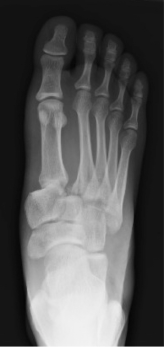
A 26 year old male with a past medical history of schizophrenia presents after jumping out of a second story window in a possible suicide attempt. He was brought to the emergency department on petition and was originally taken to the crisis center, where, in addition to intramuscular haloperidol and lorazepam, he also received a foot X-ray, which is provided below.

He has no other injuries and complains only of right foot pain. Examination shows deformity of the dorsal aspect of the right foot with tenderness and soft tissue edema over the midfoot. Range of motion in the right ankle is intact, as are peripheral pulses and neurologic function.

8.2228.22


Questions:

  1. Based on the above X-ray, which of the following is the most appropriate course of management:

A. Order more haloperidol and lorazepam because the patient is clearly malingering and there is nothing wrong with his foot.

B. Posterior mold right leg splint (with stirrups), non-weight bearing on the right leg, crutches, adequate analgesia and rapid orthopedic outpatient follow up (following completion of psychiatric evaluation)

C. Pain control, preoperative laboratory studies, and emergency department orthopedic consultation

D. Post-op shoe, pain control, PRN orthopedic or podiatric follow up.


  1. In addition to the findings that you identified on the above x-ray, which of the following other injuries is also likely present:

A. Occult talar dome fracture

B. Disruption of the ligamentous structure of the midfoot at the tarsometatarsal joint

C. Disruption of the vascular supply of the fifth metatarsal head

D. Calcaneal tendon rupture


  1. Failure to diagnose and appropriately manage this injury pattern is most commonly associated with which of the following:

A. Midfoot instability and collapse, severe arthritis

B. Avascular necrosis of the fifth metatarsal head

C. Atrophic degeneration of musculature of the dorsal foot including extensor digitorum brevis

D. Fracture non-union


Answers:

  1. C
  2. B
  3. A

Discussion:

The radiographs demonstrate a fracture through the base of the second metatarsal. In addition, there is widening of the joint space between the base of the first and second metatarsals and inferolateral subluxation of the first and second metatarsals relative to their respective cuneiforms. As with seemingly all orthopedic injuries, this pattern has an eponymous description. This injury pattern is known as a Lisfranc injury.

Lisfranc injuries refer more generally to a pattern of injury that involves disruption of the Lisfranc joint. As shown below, the Lisfranc joint is described anatomically as the articulation between the bases of the metatarsals and cuneiform bones.

This joint extends across the midfoot. Any disruption with or without fracture can be described as a Lisfranc injury. Notably, not all such injuries involve obvious metatarsal fractures; some are subtle and involve only ligamentous injury. Note that in the normal anatomical relationships of the midfoot, the proximal metatarsal articulates with the tarsal bones such that the borders of each are aligned, as shown below. The medial border of the second metatarsal aligns with the medial border of the middle cuneiform on the AP view. Presence of an avulsion fragment within the joint space between the first and second metatarsals is known as a Fleck sign (because you can never have enough eponymous ortho descriptors).

Mechanistically, Lisfranc injuries occur as a result of either direct (i.e. blunt) or indirect trauma. With an indirect traumatic injury, the Lisfranc joint undergoes excessive pronation or supination in an already plantar flexed foot, resulting in ligamentous injury. Examination will show midfoot tenderness, soft tissue swelling, potential ecchymosis, and difficulty or inability to bear weight. Plantar ecchymosis is an exam finding specific for Lisfranc injury.

Radiographic evaluation for Lisfranc injuries should focus on the relationships of the midfoot structures described above. In reviewing the x-rays in this case, note the abnormal relationship of the base of the second metatarsal to the cuneiform (red), the widening of the space between the first and second metatarsals (yellow), and the Fleck sign (blue).

Some Lisfranc injuries can be subtle. If history and exam is suggestive of more severe injury than demonstrated by x-ray, weight bearing radiographs or CT should be considered. Failure to diagnose and obtain appropriate early fixation is associated with increased complication rates.

All suspected Lisfranc injuries warrant ermegency department orthopedic consultation, and most will undergo operative fixation and extensive casting as an outpatient. Even when appropriately diagnosed and managed, there is a high degree of post-operative complications, primarily residual pain.

References:

Ross G,Cronin R,Hauzenblas J,Juliano P. Plantar ecchymosis sign: a clinical aid to diagnosis of occult Lisfranc tarsometatarsal injuries. J Orthop Trauma 1996;10(2):119–22.

Rosen’s Emergency Medicine: Concepts And Clinical Practice. Marx J, Hockberger R, Walls RM, Adams J, Rosen P. Philadelphia. Mosby

M.J. Welck et al. / Injury, Int. J. Care Injured 46 (2015) 536–541

Hatem SF. Imaging of Lisfranc injury and midfoot sprain. Radiol Clin North Am 2008;46(6):1045–60.

DeOrio M, Erickson M, Usuelli FG, Easley M. Lisfranc injuries in sport. Foot Ankle Clin 2009;14(2):169–86.vier, 2010

Senior Report 8.21

seniorreport

Case Presentation by Dr. Sean Michael, MD

Visual Stimulus Case:

A 59-year-old man with COPD presents with acute dyspnea. His breath sounds are nearly inaudible. He is tripoding with accessory muscle use and suprasternal retractions. Temperature is 37.7°C, heart rate 112, respiratory rate 36, blood pressure 168/92, and oxygen saturation 89% on 2 liters via nasal cannula. Glucocorticoids and nebulized bronchodilators are administered. Bedside thoracic ultrasound is performed and demonstrates the following M-mode image in the right second intercostal space:

8.21

Questions: 

  1. The most likely etiology of the ultrasonographic finding above is:

A. Emphysematous bulla or apical bleb

B. Iatrogenic pneumothorax

C. Lobar pneumonia

D. Primary spontaneous pneumothorax


Additional images are obtained of the right chest at the level of the fifth intercostal space:


8.211


  1. Given the new information obtained in this image, which of the following is the best course of action:

A. CT Thorax

B. Intravenous antibiotics

C. Non-invasive positive pressure ventilation

D. Tube thoracostomy


  1. The findings in the second ultrasound image serve mostly to:

A. Increase diagnostic sensitivity (ie. have a high negative predictive value)

B. Increase diagnostic specificity (ie. have a high positive predictive value)

C. Predict a decreased risk of mortality

D. Predict an increased risk of treatment failure


Answers and explanation:

1. A
2. D
3. B

(Note: This explanation assumes that you understand the basics of lung ultrasound. If you need a refresher, there are lots of great online resources.)

This patient presented with an apparent moderate to severe COPD exacerbation with hypoxia and poor air exchange on lung auscultation. Bedside lung ultrasound (image 1) demonstrates absence of pleural sliding on M-mode. In the setting of COPD (or many other critical illness states), the absence of lung sliding may be caused by any number of pathophysiologic conditions. While the absence of lung sliding is quite sensitive for pneumothorax of any etiology, in comparison to patients with traumatic pneumothorax, patients with non-traumatic dyspnea may have numerous other causes of poor lung sliding, which may increase the false-positive rate for ultrasound exams (Slater 2006, Lichtenstein 2008).

In this clinical presentation, the most likely reason for the absence of lung sliding is an emphysematous bulla/apical bleb (question 1, answer A). Bullae are common in COPD, especially in advanced disease, and a ruptured apical bleb is a common cause of secondary spontaneous pneumothorax (Noppen 2008). While iatrogenic pneumothorax (question 1, answer B) is a known complication of a number of procedures, this patient did not undergo any high-risk interventions, such as transthoracic needle aspiration, central venous access, thoracentesis, transbronchial or pleural biopsy, or positive pressure ventilation (Sassoon 1992). Lobar pneumonia (question 1, answer C) on ultrasound is characterized mostly by an A-B profile or by the absence of lung sliding with a B profile (Lichtenstein 2008). Primary spontaneous pneumothorax (question 1, answer D) typically occurs in young male smokers with thin body habitus and (by definition) is not secondary to underlying pulmonary disease, such as COPD, cystic fibrosis, or malignancy (Noppen 2008).

The second ultrasound image shows a lung point, which is much more specific for pneumothorax (Lichtenstein 2008). Given the patient’s dyspnea, hypoxia, and the size of the pneumothorax (from at least the second through the fifth intercostal spaces, but more likely from the apex through the fifth), the correct intervention is tube thoracostomy (question 2, answer D). This can be accomplished with either a small bore surgical chest tube or via percutaneous small-bore catheter (aka “a pigtail”) (Contou 2012, Tsai 2006). There is already enough clinical information to diagnose pneumothorax, and CT Thorax (question 2, answer A) is not required. Intravenous antibiotics (question 2, answer B) might be indicated in suspected bacterial pneumonia, but the second ultrasound image is not diagnostic of pneumonia. Non-invasive positive pressure ventilation (question 2, answer C) may be required for this patient, which is an even more compelling reason to perform thoracostomy. The ultrasound does not predict need for NIPPV, however.

As mentioned previously, lung point dramatically increases specificity (question 3, answer B) for pneumothorax (Lichtenstein 2008 and lots of other papers—this isn’t a comprehensive review). The most sensitive (question 3, answer A) finding is absence of lung sliding (or perhaps absence of sliding with augmented color power Doppler) (Cunningham 2002, Lichtenstein 2008, etc). Lung ultrasound has not yet been shown to predict either treatment failure (question 3, answer C) or mortality (question 3, answer D) in the setting of secondary spontaneous pneumothorax.

In this case, a small-bore chest tube was placed, the patient placed on bi-level NIPPV, and he did well.

References:

Contou D, Razazi K, Katsahian S, et al. Small-bore catheter versus chest tube drainage for pneumothorax. American Journal of Emergency Medicine. 2012;30(8):1407–1413. doi:10.1016/j.ajem.2011.10.014.
Cunningham J, Kirkpatrick AW, Nicolaou S, et al. Enhanced recognition of “lung sliding” with power color Doppler imaging in the diagnosis of pneumothorax. J Trauma. 2002;52(4):769–771.
Lichtenstein DA. Relevance of Lung Ultrasound in the Diagnosis of Acute Respiratory Failure*. Chest. 2008;134(1):117. doi:10.1378/chest.07-2800.
Noppen M, De Keukeleire T. Pneumothorax. Respiration. 2008;76(2):121–127. doi:10.1159/000135932.
Sassoon CS, Light RW, OHara VS, Moritz TE. Iatrogenic pneumothorax: etiology and morbidity. Respiration. 1992;4:215–20.
Slater A, Goodwin M, Anderson KE, Gleeson FV. COPD can mimic the appearance of pneumothorax on thoracic ultrasound. Chest. 2006;129(3):545–550. doi:10.1378/chest.129.3.545.
Tsai W-K, Chen W, Lee J-C, et al. Pigtail catheters vs large-bore chest tubes for management of secondary spontaneous pneumothoraces in adults. The American journal of emergency medicine. 2006;24(7):795–800. doi:10.1016/j.ajem.2006.04.006.
Ultrasound for Detection of Pneumothorax. Rebel EM (http://rebelem.com/ultrasound-detection-pneumothorax). Accessed 3/20/2015.

Intern Report 8.20

internreport

Case Presentation by Khoa Nguyen, MD

CC: “fever and runny nose”


HPI: 9-day-old full term female born via C-section who presents with cough, rhinorrhea, and tactile fever. Patient’s mother stated that the patient had rhinorrhea 3 days ago who then developed a cough the following day. The mother then felt that the patient was warm in the back but did not measure any temperature. The patient was not given any anti-pyretic or antibiotics. There had been green discharges from the eyes. Patient had 2 episodes of non-bloody nonbilious emesis that looked like her feeds. Patient had been sleeping more than normal. On further questioning, the patient’s mother had GBS and Chlamydia with this pregnancy and had HSV during the previous pregnancy.   There were no changes in the number of wet diapers and no changes in PO intake.


ROS:

Constitutional: positive for tactile fever

HEENT: positive for rhinorrhea, green discharges from eyes, and congestion

CV: neg

Pulmonary: positive for coughing

GI: positive for 2 episodes of NBNB emesis. No changes in appetite and PO intake.

GU: no change in number of wet diapers

The rest of the ROS were negative


PMH/PSH: none

Allergies: NKDA

Immunizations: UTD

Birth history: 39 wks, repeat c-section

Family history: Mother was treated for GBS and chlamydia with this pregnancy


Physical Exam:

Vital signs: Temperature 37, HR 160, RR 30, BP 67/41, 98% on RA

General: patient is alert and responsive to touch

HEENT: NC/AT, anterior fontanelle is open, soft, and flat. There is bilateral eye discharge with crusting. No chemosis. Eyelids appear normal. TMs are clear. Oropharynx within normal limits

Neck: supple

CV: RRR, S1 S2, no notable murmurs

Lung: Clear to auscultation bilaterally

GI: soft, nontender, non distended, no masses

MSK: moving all extremities

Skin: no rashes, bruising

Neuro: normal moro, rooting, grasp


Questions:

  1. What is the workup for this patient?

A. Patient does not need a workup.

B. Full sepsis workup: LP, CBC, CXR, LP, UA with culture, blood culture, RSV/Flu.

C. UA with culture, CBC, Chest x-ray

D. Rapid Viral antigen testing


  1. What is the management?

A. PO challenge and discharge home after reassuring the mother that this is likely a viral infection and that she needs to follow-up with PMD.

B. Ampicillin, ceftriaxone, and acyclovir

C. Ampicillin and acyclovir

D. Ampicillin, cefotaxime, and acyclovir


  1. Which of the following is true?

A. Management of pediatric fever is the same throughout all ages.

B. Defervescence after acetaminophen administration has been shown to reliably exclude bacteremia in children of any ages.

C. The absence of fever does not eliminate the possibility of serious bacterial illness.

D. A thorough history and physical exam can exclude a serious bacterial illness in a patient less than 28 days old.


Answers:

1. B
2. D
3. C


Pediatric patients from 0-28 days of age who present with a fever are at a high risk for bacterial illness. Fever may be the only clinical manifestation of a potentially life-threatening disease. However, in this age group, the absence of fever does NOT eliminate the possibility of serious bacterial illness because more than half of neonates with meningitis are afebrile.

The physical exam in this age group is insensitive to exclude serious bacterial illness.

Here are some of the findings to suggest bacterial meningitis:

Vital signs – apnea, tachypnea, hypothermia, hyperthermia, bradycardia, tachycardia. The absence of fever does not rule out the possibility of serious bacterial illness.
Behavior – listless, restless, irritable, lethargy, change in sleeping pattern
Neurologic – high pitch cry, nystagmus, vacant stare, seizure, altered tone, absence of cry
Dermatologic – cyanosis, petechiae, purpura, livedo reticularis
GI – altered feeding, diarrhea, vomiting, abdominal distention, jaundice


B – Although the patient has symptoms to suggest a viral infection, she needs a thorough workup given her age. A change in sleeping pattern, cough, eye discharges, and tactile fever must be taken seriously. Additionally, the patient’s mother has a history of GBS, chlamydia, and HSV which all put the patient at risk of a serious infection. For these reasons, the patient needs a complete sepsis evaluation.
D – The patient should be treated empirically with broad spectrum medications in the ED given her risks of a serious infection. Ceftriaxone should be avoided in patients younger than 28 days because of a hypothetical risk of causing bilirubin encephalopathy since this medication causes bilirubin to be displaced from protein binding sites.
C – Management of pediatric fever depends on the age of the patient. Defervescence of a fever after Tylenol ingestion does not exclude bacteremia in children of any ages.


References:

Marx, JA, Hockerberger R, Walls RM. Rosen’s Emergency Medicine: Concepts and Clinical Practice (8th edition), Mosby 2013.

Senior Report 8.19

seniorreport

 Case Presentation by Laura Smylie, MD

A 14 year old girl who presents with nausea and vomiting for one day and an abrupt onset of chest pain.
Vitals: BP 100/67, HR 121, RR 25, Temp 36.7, 100% on room air.

1

2


Questions:
1. What is the diagnosis based upon the above x-rays?
a) Foreign body
b) Pneumothorax
c)Pneumomediastinum
d)Apical pneumonia

2. What is the likely etiology of the radiographic finding?
a) alveolar rupture secondary to forceful retching
b) esophageal rupture secondary to forceful retching
c) acute PE
d) no abnormality on chest x ray.

3. What is the most appropriate initial management of this patient?
a) place on continuous pulse oximetry, place a left sided chest tube
b) place on continuous pulse oximetry, 4mg ondansetron, NPO.
c) place on a continuous cardiac monitor, start heparin drip, consult cardiology.
d) send blood cultures, start antibiotics, encourage PO intake.


Answers and discussion:

1) C
2) A
3) B

1) C – Pneumomediastinum. You can see air tracking in the soft tissues of the neck and the upper aspects of the mediastinum. Although there could potentially be a pneumothorax (B) associated with this this, no pneumothorax can be seen in this study. No foreign body (A) is present and there is no infiltrate to suggest pneumonia (D).

1


2) A – Alveolar rupture secondary to forceful retching is the most likely etiology of the pneumomediastinum, although you should also be concerned for possible esophageal rupture secondary to forceful wretching (B) also known as Boerhaave’s esophagus. Review of the literature shows that in similar presentations, esophograms are typically negative for tears in the esophagus. Acute PE (C) has not been shown to present with free air. On a chest xray, the most concerning (and classically pimped) findings for acute PE are Hampton’s Hump and Westermark’s sign. Hampton’s Hump, represented in the first image below, shows a wedge shaped area of hyperdensity along the lung parenchyma periphery, indicative of an infact/PE. Westermark’s sign, as shown in the second image below, shows a focal peripheral hyperlucency secondary to oligemia, with or without dilation of the central pulmonary vessels.

2


3) B – Although there is no obvious pneumothorax on the initial chest x ray, you must keep a high level of suspicion for a small pneumothorax. This would not necessitate chest tube placement (A), but a nonrebreather and continuous pulse oximetry are appropriate if a small pneumothorax is present. Given that the retching led to the pneumomediastinum, treat her nausea with ondansetron. She should be kept NPO until an esophagram can be obtained (as an inpatient or in the observation unit) to definitively rule out Boerhaave’s esophagus. As an inpatient, the chest x ray should repeated in 6-8 hours. C is the treatment for a non-massive PE or NSTEMI; D is the treatment for pneumonia, neither of which applies in this case.

3

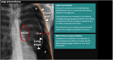
The percentage of pneumothorax will guide therapy. This picture illustrates that 2 cm pneumo is typically the cut off point for inserting a chest tube with a spontaneous pneumo but not necessarily with a traumatic pneumo.

4


References:

Spontaneous pneumomediastinum: diagnostic and therapeutic interventions. Al-Mufarrej F, Badar J, Gharagozloo F, Tempesta B, Strother E and Margolis M. Journal of Cardiothoracic Surgery 2008, 3:59 doi:10.1186/1749-8090-3-59

BMJ Case Rep. 2012 Oct 10;2012. pii: bcr0320091647. doi: 10.1136/bcr.03.2009.1647.

http://radiopaedia.org/articles/westermark-sign

Gantner J, Keffeler JE, Derr C. Pulmonary embolism: An abdominal pain masquerader. J Emerg Trauma Shock [serial online] 2013 [cited 2015 Mar 26];6:280-2. Available from: http://www.onlinejets.org/text.asp?2013/6/4/280/120376