Case Presentation by Dr. Daniel Hutchens, MD
History:
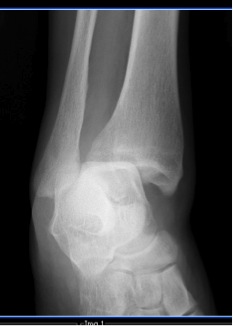
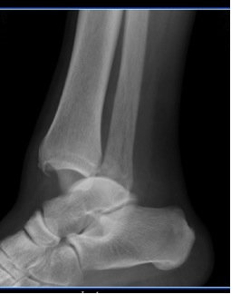
66 year old female presents with right ankle pain after slipping on a mat and twisting it. She was unable to bear weight on it immediately after the fall. She noticed immediate pain and swelling. She denies pain in any other joints. She denies any loss of consciousness. She has no other complaints.
Physical Exam:
Cardiovascular: Regular rate and rhythm, no murmurs, no S3/S4, radial and dorsalis pedis pulses present and equal bilaterally in both upper and lower extremities.
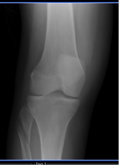
Musculoskeletal: Obvious deformity of the right ankle. Decreased range of motion in the right ankle when compared to the left. Tenderness to palpation over the right medial malleolus. No tenderness to palpation over the distal tibia or fibula. Mild tenderness to palpation over the right fibular head.
Neurologic: Alert and oriented to person, place, and time. Smile symmetric, tongue protrudes midline, uvula raises midline, eyebrows raise symmetrically, eyes close with equal strength. Sensation to light touch equal and intact in bilaterally lower extremities.
Questions
- What musculoskeletal physical exam points must be covered in a patient with traumatic ankle pain?
a. Assessment for deformity, range of motion, palpation of inferior and posterior edges of medial/lateral malleoli, and first 6 inches of fibula and tibia.
b. Assessment for deformity, range of motion, palpation of inferior and posterior edges of medial/lateral malleoli, first 6 inches of fibula and tibia, and calcaneous.
c. Assessment for deformity, range of motion, palpation of medial/lateral collateral ligaments, syndesmotic ligaments, inferior and posterior edges of medial/lateral malleoli, entire length of fibula and tibia.
d. Assessment for deformity, range of motion, palpation of medial/lateral collateral ligaments/syndesmotic ligaments, inferior and posterior edges of medial/lateral malleoli, entire length of fibula and tibia, base of the 5th metatarsal, and calcaneous. - What is your radiologic diagnosis?
a. Pott’s fracture
b. Maisonneuve fracture
c. Cotton fracture
d. Dupuytren’s fracture - What is the best disposition of this patient with this type of fracture?
a. Walking boot with orthopedic follow-up in 2 weeks.
b. Surgical repair of the ankle with intramedullary rod placement in the fibula.
c. Surgical repair of the ankle, non-weight-bearing status for 9-12 weeks.
d. Ankle reduction in the ER, non-weight-bearing status, orthopedic follow-up in 6 weeks.
Answers & Discussion
1) d
2) b
3) c
1. Patient’s presenting with traumatic ankle pain carry a differential from ranging from very benign to severe. Physical examination should begin with assessment of deformity, ecchymosis, edema, and perfusion.
Palpation should include:
- medial and lateral malleoli
- syndesmotic ligaments
- inferior and posterior edges of the medial and lateral malleoli
- entire length of the tibia and fibula
- medial and lateral dome of the talus
- base of the fifth metatarsal, the calcaneous
- Achilles tendon
- peroneal tendons behind the lateral malleolus
Evaluation of weight bearing should be performed only if suspicion for fracture is low. A useful clinical decision rule to risk stratify patients with traumatic ankle pain was developed in 1993. The Ottawa Ankle Rules were developed to aid in the efficient use of radiography in acute ankle and mid-foot injuries. The original article was published in JAMA, titled “Decision Rules for the use of Radiography in Acute Ankle Injuries.” The rules have since been prospectively validated on multiple occasions in different populations and in both children and adults with sensitivity approaching 100%.
2. Classic definition of a Maisonneuve fracture is medial ankle disruption (deltoid ligament tear or medial malleolar fracture) with complete tearing of the syndesmotic ligament joining the tibia and fibula, and fracture of the proximal fibula. The injury results from an external rotation force that transmits through the syndesmotic ligament, tearing it, and exits through the proximal fibula, fracturing it. A Dupuytren’s fracture is similar to a Maisonneuve and synonymous to a Pott’s fracture. Instead of a proximal fibular fracture it involves a distal fibular fracture with dislocation of the ankle and disruption of the syndesmotic ligament. A Cotton’s fracture is a trimalleolar fracture that involves the lateral malleolus, medial malleolus, and distal posterior aspect of the tibia (posterior malleolus).
3. Whether to admit or discharge with orthopedic follow up depends on location of the ED. If orthopedics is available in hospital then consultation should be obtained for consideration of operative management. Admission for surgery should be discussed but is not emergently necessary. The patient does need to be splinted (sugar tong & posterior mold) and should strict non-weight bearing. If Orthopedics consultations is not available within the ED then they should be contacted by phone to arrange close follow up with in the next several days. Maisonneuve fractures are unstable ankle fractures. For an ankle fracture to be unstable the restraining structures on the inside of the ankle (deltoid ligament or medial malleolus) are disrupted. These fractures require surgery to re-stabilize the syndesmotic ligament and ankle mortise. Complications include peroneal nerve injury as with any fracture of the proximal fibula, as well as ankle arthrosis and compound injury. Time to recovery post-operatively is usually 6 weeks in a short leg non-weight bearing cast.
References:
- Marx, John A, et al. Rosen’s Emergency Medicine, 7th Ed. Philadelphia: Mosby Elsevier, 2010. Print.
- Wheeless, Clifford R. “Maisonnueve Fracture.” Wheeless’ Textbook of Orthopedics. N.p., 22 May 2012. Web. 26 Oct. 2014.
- Kalyani, Bharati S, et al. “The Maisonnueve Injury: A Comprehensive Review.” Healio Orthopedics 3.33 (2010): n. pag. Web. 26 May 2014.
Filed under: Senior Report, Uncategorized | Tagged: ankle pain | 2 Comments »


